
Ventricular Septal Defect
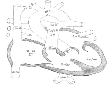
Definition: interventricular septal communication (IVS) defined by its location. The IVS can be divided into a muscular and membranous portion, hence perimembranous VSD (80%) or muscular VSD (inlet/trabecular/outlet = infundibular region). It is the most common type of CHD.
Incidence: 1.7-53:1000 of all live births.
Prevalence: 20% as a single lesion, 50% as part of lesion in CHD.
Physiology:
Depends on the size, PVR, RVP and LVP
- in neonates with increased PVR there is minimal shunting
- where there is a drop in PVR and and therefore less restrictive, there is a left to right shunt leading to volume load in the LA and LV. This can lead to CCF and, if long standing, Eisenmenger syndrome
Diagnosis:
- Clinical examination: auscultation – holosystolic murmur; cyanosis; and clubbing in Eisenmenger syndrome
- ECG: LVH
- CXR: increased pulmonary blood blow (in Eisenmenger there is reduced PBF)
- ECHO: for size, location, shunt, haemodynamic evaluation
- MRI: limited usefulness
- Cardiac catheterization: PVR, response to pulmonary vasodilators, Qp:Qs
Preoperative management:
Treat CCF: CPAP/IPPV if required; digoxin; frusemide (1mg/kg up to QID); spironolactone (1mg/kg BD).
Aim for an afterload reduction:milrinone (0.25-0.5 mcg/kg/min) or captopril (0.1-2 mg/kg TDS).
Preoperative preparation:
ECG, CXR, CUS, FBE, clotting, UEC, PRBC (4), FFP (2), platelets (2), cryoprecipitate (2). methylprednisolone 10mg/kg 12hrs and 6hrs pre surgery in neonates.
Surgery:
Pulmonary artery banding for protection of pulmonary circulation in neonates with multiple VSDs and CCF
In children >3 months (Qp:Qs >1.5:1), surgical closure via transatrial approach, sometimes by right ventriculotomy, and rarely by left apical ventriculotomy. Catheter closure by amplatzer device. Heart-lung transplantation in Eisenmenger syndrome.
Postoperative management:
- Inotropes: milrinone plus dopamine or adrenaline (plus noradrenaline). Aim for MAP >40 mmHg in neonates.
- Haemodynamics: age adjusted (in neonates: SBP >60 mmHg, MAP >40 mmHg, CVP 8-12 mmHg).
- Respiratory: normoxaemia, normocapnea.
- Fluid restriction: 1ml/kg/hr, early feeds.
- Continue preoperative diuretic therapy.
- Haemostasis.
- If catheter closure: aspirin 5mg/kg OD once feeds tolerated.
Specific problems:
- Risk of pulmonary hypertension in older children and Qp:Qs >1.5:1.
- Arrhythmia: complete heart block, SVT, JET.
Outcome:
Average ICU stay: 2 days.
Mortality: <1% in isolated VSD, but up 5-10% in multiple defects.
Residual shunts: 30% after surgical closure, majority close spontaneously.
References:
[1] Br Heart J. 1980 Mar;43(3):332-43: Soto et al: Classification of ventricular septal defects
[2] Circulation. 2006 Nov 14;114(20):2190-7: Minette et al: Ventricular septal defects
[3] Cardiol Young. 2007 Jun;17(3):243-53: Butera et al: Percutaneous closure of ventricular septal defects
All Marc’s PICU cardiology FOAM can be found on PICU Doctor and can be downloaded as a handy app for free on iPhone or Android. A list of contributors can be seen here.


